Fig. 2.
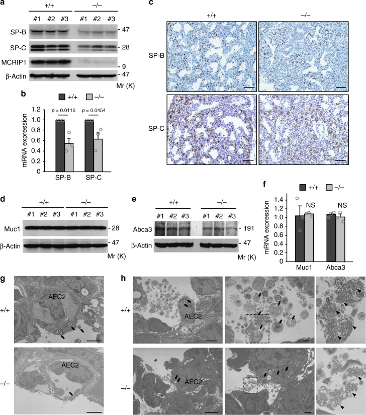
Surfactant proteins are down-regulated in Mcrip1-KO mouse lungs. a, b Immunoblotting a and qRT-PCR b analyses of SP-B and SP-C expression in whole-lung lysates isolated from Mcrip1+/+ and Mcrip1−/− mice. Data in b represent the mean ± SEM. obtained from three mice. c Immunohistochemical analyses of SP-B and SP-C expression in the lung tissues of Mcrip1+/+ and Mcrip1−/− neonates. Scale bars, 100 μm. d–f Immunoblot d, e and qRT-PCR f analyses of the expression of Muc1 and Abca3 in whole-lung lysates isolated from Mcrip1+/+ and Mcrip1−/− mice. Data in f represent the mean ± SEM obtained from three mice. NS, not significant. g, h Ultrastructure of AEC2, lamellar bodies, and tubular myelin in Mcrip1+/+ and Mcrip1−/− lungs at E21 g and at P0 h. h The areas in the small squares in the middle sets of panels were enlarged and are shown on the right. Arrows indicate lamellar bodies and arrowheads indicate tubular myelin. Scale bars, 2 μm in g. Scale bars, 2 and 1 μm in left and middle panels of h, respectively

