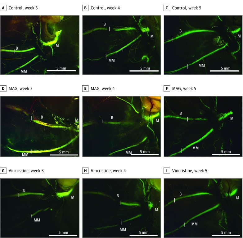
Figure 2. Representative Images of Facial Nerve With Buccal and Marginal Branches Visible.
Nerve regeneration is demonstrated for the saline control group at weeks 3 (A), 4 (B), and 5 (C); the myelin-associated glycoprotein (MAG) group at weeks 3 (D), 4 (E), and 5 (F); and the vincristine group at weeks 3 (G), 4 (H), and 5 (I). Images were taken at × 1.25 magnification (scale bars represent 5 mm). The branches of the nerve are identified as main trunk (labeled M), buccal (labeled B), and marginal mandibular (labeled MM). Sites of injury are denoted by a white bar placed alongside the appropriate segment of each proximal nerve branch.

