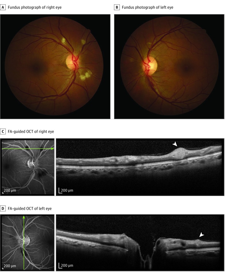
Figure 1. Images From Patient With Bilateral Retinopathy and Confirmed Yellow Fever Admitted to Intensive Care Unit Because of Systemic Decompensation With Hepatic and Renal Failure, Encephalopathy, and Profuse Gastrointestinal Bleeding.
Fundus photographs show retinal nerve fiber layer (RNFL) infarcts (A and B); a flame-shaped hemorrhage is present in the inferotemporal arcade of left eye (B). Fluorescein angiography (FA) discloses hypofluorescent blockage at the level of RNFL infarcts, with no evidence of hyperfluorescence of the optic disc, macula, or retinal blood vessels (C and D). Fluorescein angiography–guided optical coherence tomography (OCT) (C and D) shows hyperreflective nodules at the RNFL (white arrowheads) corresponding to the RNFL infarcts. Green lines indicate retinal topography of OCT section.

