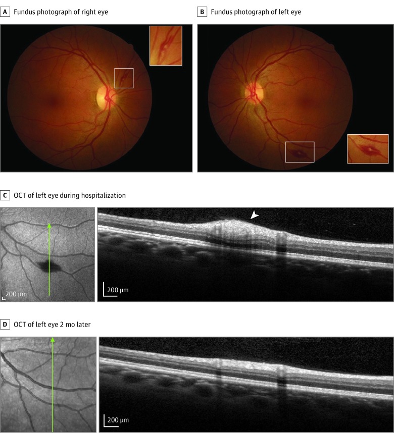
Figure 2. Images From Patient With Bilateral Retinopathy Admitted to Intensive Care Unit Because of Systemic Decompensation With Renal Failure, Thrombocytopenia, and Coagulopathy.
Fundus photographs show superficial hemorrhages (white rectangles; A and B), including a Roth spot in left eye (B). Insets are magnifications of the areas shown in the white rectangles. Optical coherence tomography (OCT) of the left eye over the Roth spot (white arrowhead) is shown during hospitalization (C) and 2 months later, after recovery (D). Green lines indicate retinal topography of OCT section.

