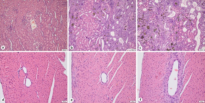
Fig. 6.
Histopathology. Hematoxylin-eosin stains of kidney (a–c) and right heart ventricle (d–f) of control (a, d), AD-2K (b, e) and AD-1K (c, f) rats. b, c Brownish adenine crystals with giant cell formation, cystic tubular dilatation, interstitial fibrosis, mononuclear cell infiltrate, and tubular atrophy/degeneration. The semiquantitative degree of tubular damage was marked in AD-1K rats (c), while tubular damage in AD-2K rats (b) was only moderate, with intact eosinophilic areas (left half of the picture). Vasculopathy of slight degree with thickening of the ventricular artery wall was only seen in AD-1K rats (f). Scale bars, 100 μm. AD-1K rats, rats with unilateral nephrectomy, adenine at 0.75% in the diet; AD-2K rats, sham-operated rats, adenine at 0.75% in the diet.

