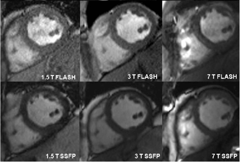
FIGURE 10.

Short axis cardiac images acquired at 1.5, 3, and 7 T using fast low angle shot (FLASH) and steady state free precession (SSFP) sequences in the same subject. Republished with permission of John Wiley and Sons, from “7 Tesla (T) human cardiovascular magnetic resonance imaging using FLASH and SSFP to assess cardiac function: validation against 1.5 T and 3 T,” Suttie JJ, Delabarre L, Pitcher A, et al. NMR in Biomedicine. 2012;25(1).
