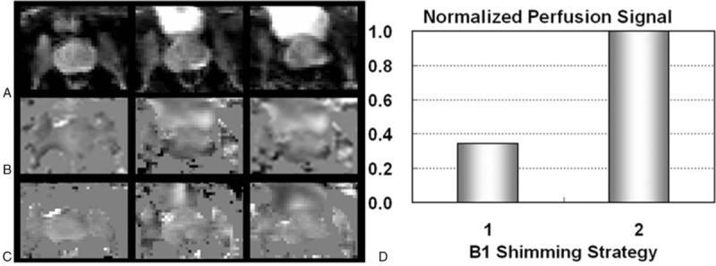
FIGURE 17.

Prostate perfusion imaging results from a subject acquired at 7T. Shown are proton density images at three locations (A) followed by perfusion-weighted imaging maps acquired with a fixed single B1+ shimming solution (B) and dynamically applied multiple B1+ shimming solutions (C). (D) Normalized prostate perfusion signals are presented using the two B1+ shimming strategies: strategy 1) using a single B1+ shim optimized for efficiency on the prostate, and strategy 2) using multiple dynamically applied B1 shimming solutions for different RF components.
