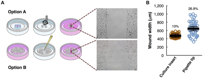
Figure 1.
Overview of the wound healing assay preparation protocols. (A) Step-by-step scheme showing the differences between wound healing protocol using a culture insert (option A) and using pipette tip (option B). Phase-contrast microscopy shows gap appearance and both cell fronts just before to start the time-lapse experiment. (B) Measurements of wound width (μm) in culture insert (n = 50) or pipette tip (n = 50). Mean values (thick horizontal lines), confidence limits (α = 0.05, thin horizontal lines), and coefficients of variation (label) are shown.

