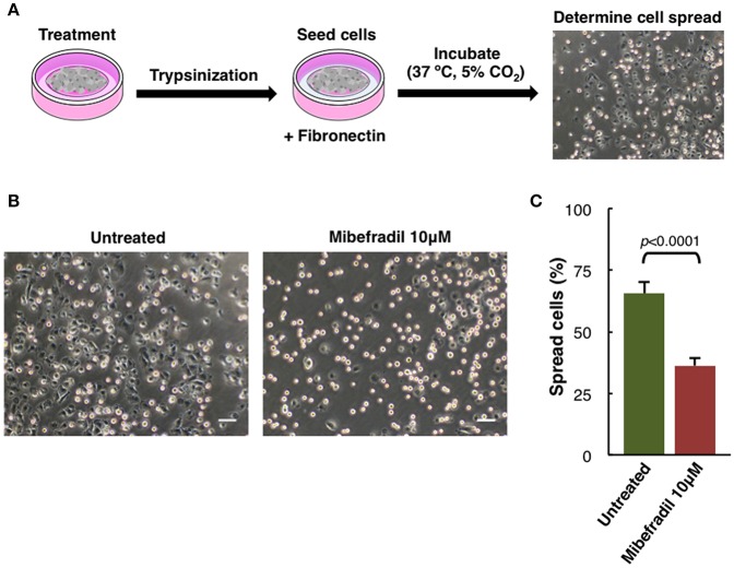
Figure 5.
Cell spreading assay in M3 melanoma cells. (A) Experimental design scheme of spreading assay. Cells were treated for 24 h, trypsinized and seeded onto fibronectin (10 μg ml−1) coated plate. After 1 h cells were fixed with 2% PFA and (B) phase-contrast images were captured. Scale bars, 50 μm. (C) Plot showing the percentage of spreading cells. Round bright cells were considered unspread. Values are percentage of spread cells ± SD (n = 3 independent experiments; at least 600 cells for each experiment were counted). The corresponding p-value obtained by unpaired two-tailed Student's t-test is shown.

