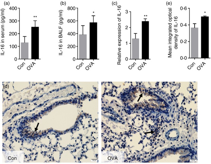
Figure 2.

Levels of interleukin‐16 (IL‐16) in serum, bronchoalveolar lavage fluid (BALF), and lung tissue are elevated in C57BL/6 mice upon ovalbumin (OVA) ‐induced allergic inflammation. ELISA was used to detect the expression level of IL‐16 in serum (a) and BALF (b). The mRNA expression of IL‐16 in lung (c). IL‐16 protein expression in the paraffin section of lung was detected by Immunohistochemistry (Magnification: 100×) (d and e). The mean integrated optical density (IOD)was semiquantified using image‐proplus software. Date are expression as mean ± SEM (n ≥ 5). *P < 0·05 and **P < 0·01 versus control (Con).
