Figure 1.
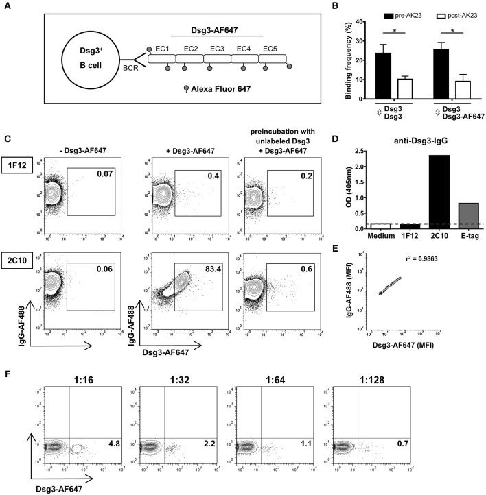
Detection of Dsg3-specific B cell hybridoma (BCH) using fluorescently labeled Dsg3. (A) Schematic drawing: the recombinant human extracellular domain (EC1-EC5) of Dsg3 was labeled with the fluorescent dye Alexa Fluor 647 (Dsg3-AF647) and was used for staining of Dsg3-specifc B cell receptors (BCR). (B) Binding of Dsg3-AF647 to Dsg3 ± addition of the monoclonal Dsg3-specific antibody AK23 was evaluated with atomic force microscopy. Cumulative data from 3 individual measurements with five replicates for each condition are presented as mean + SD. Statistical analysis was performed by multiple t-tests followed by Šidák correction. Differences between groups were considered statistically significant at p-values of <0.05 indicated as *. (C) Binding efficacy of Dsg3-AF647 to Dsg3-specific BCR was determined by staining of a Dsg3-specific BCH (2C10) and an unrelated BCH (1F12) together with anti-IgG antibody. Binding of Dsg3-AF647 was blocked by preincubation with unlabeled Dsg3. FACS plots shown are representative of three individual experiments. (D) Specificity of monoclonal BCH cells for Dsg3 was tested by ELISA. Anti-E-Tag served as positive and culture medium as negative control. (E) Correlation of mean fluorescence intensity (MFI) of Dsg3-AF647 with surface IgG. (F) Titration of Dsg3-specific BCH (2C10) cells in unrelated 1F12 cells in a calculated ratio of 1:16 (6.25%), 1:32 (3.13%), 1:64 (1.56%), and 1:128 (0.78%) representative of three individual experiments.

