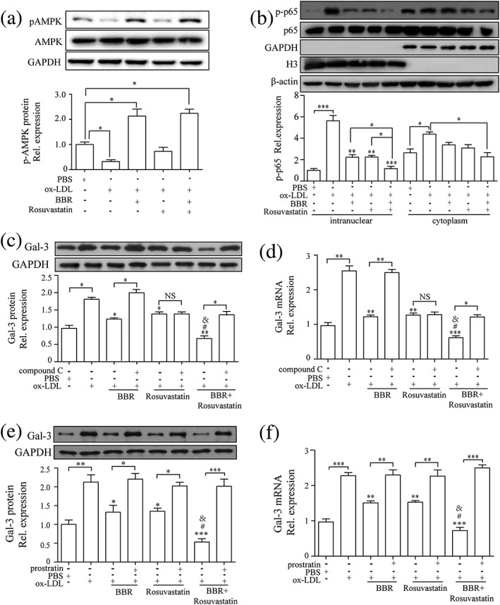
Figure 6.

Berberine (BBR) downregulated galectin‐3 expression by inhibiting NF‐κB and activating AMPK signaling pathway. (a–d) Macrophages were pretreated with phosphate‐buffered saline (PBS), BBR (25 μM), rosuvastatin (25 μM), and combination of BBR (25 μM) and rosuvastatin (25 μM) for 1 hr and then stimulated by 100 μg/ml oxidized low‐density lipoprotein (ox‐LDL) for 30 min. Western blots and quantification of (a) AMPK and phospho‐AMPK (p‐AMPK) and (b) NF‐κB p65 and phospho‐NF‐κB p65 (p‐p65). Protein and mRNA expression of galectin‐3 measured by Western blotting and real‐time PCR on THP‐1‐derived macrophages pretreated with PBS, BBR, rosuvastatin, and BBR and rosuvastatin, in the presence or absence of (c and d) compound C (10 μg/ml) or (e and f) prostratin (10 μg/ml) for 1 hr before the induction by 100 μg/ml ox‐LDL for 24 hr. Galectin‐3 was normalized by GAPDH levels. Phospho‐AMPK (Thr172) was normalized by total AMPK. p‐p65 (Ser536) was normalized by NF‐κB p65. GAPDH was the indicator for cytoplasm protein. Histone H3 was the indicator for nuclear protein. β‐Actin was the indicator for both cytoplasm protein and nuclear protein. Data are represented as mean ± SD. n ≥ 3. * p < 0.05, ** p < 0.01, *** p < 0.01 versus ox‐LDL group; # p < 0.05 versus ox‐LDL + BBR group; & p < 0.05 versus ox‐LDL + rosuvastatin group. NS: non‐significant
