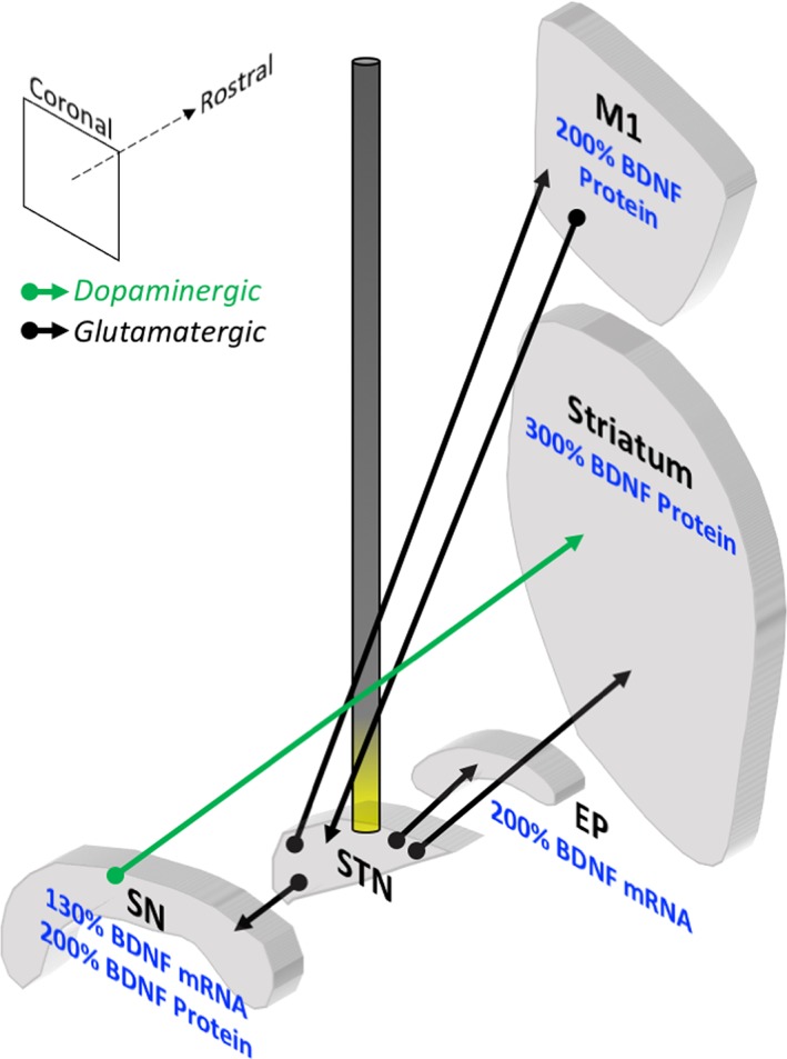
Figure 2.

STN DBS increases BDNF in the basal ganglia in PD animal models. Coronal sections of select basal ganglia structures in the rat are depicted in 3 dimensions relative to one another, and an electrode stimulating the STN is illustrated. Effects of high‐frequency stimulation of the STN on BDNF levels in the rat are noted. STN DBS increases BDNF mRNA in the SN and entopeduncular nucleus (EP, rodent homologue to primate GPi). STN DBS also increases BDNF protein in the primary motor cortex (M1) and the striatum of unlesioned animals and the SN of lesioned animals. The green arrow represents dopaminergic fibers; the black arrows represent glutamatergic fibers. Data summarized from Spieles‐Engemann et al (2011).8
