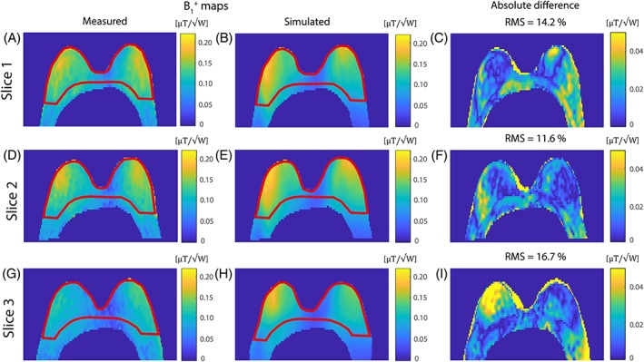
Figure 5.

Three transversal slices of the measured B 1 + map (same volunteer as ‘small’ in Figure 4) compared with the simulated B 1 + map with the same shim settings, resulting in the absolute difference map with a mean RMS of 14.2%. The measured B 1 + map was acquired with the DREAM method (T R = 7.0 ms; T E = 1.97 ms and 2.4 ms; flip angle = 10° and 60°, FOV = 320 × 520 × 60 mm3 with a resolution of 3.0 × 5.0 × 6.0 mm3 and a nominal B 1 of 12 μT). The mean B 1 + has been measured in two ROIs in the measured B 1 map, where ROI1 includes both breasts to the pectoralis muscle boundary (shown in red) and ROI2 has been set for the entire FOV shown (including the pectoral muscle and reaching into the axilla). Within ROI1 a mean B 1 + of 0.12 μT/√W, coefficient of variation 20%, was measured, and in ROI2 a mean B 1 + of 0.11 μT/√W, coefficient of variation 27%
