
Figure 4.
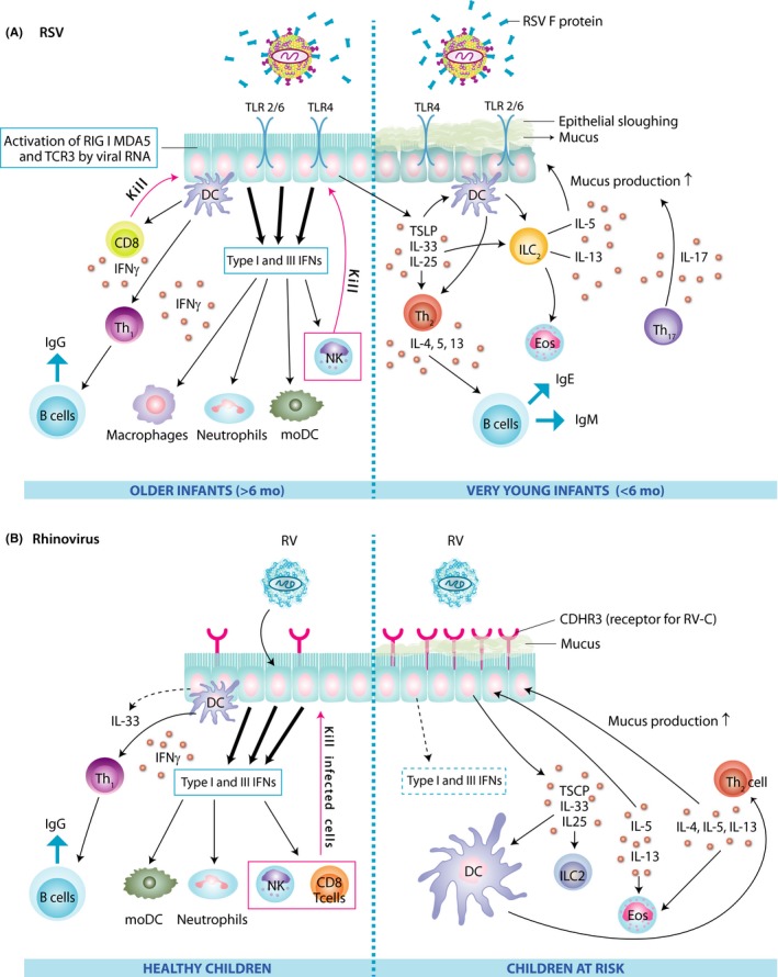
Pathogenesis of respiratory syncytial virus (A) and rhinovirus infection (B) in the airway epithelial cells of healthy children and those at risk. CDHR3, cadherin‐related family member 3; DC, dendritic cell; Eos, eosinophil; IFN, interferon; Ig, immunoglobulin; IL, interleukin; ILC, innate lymphoid cell; MDA, melanoma differentiation‐associated protein; moDC, monocyte‐derived dendritic cells; NK, natural killer cell; RIG, retinoic acid‐inducible gene; RSV, respiratory syncytial virus; RV, rhinovirus; TCE3, third T‐cell receptor; Th, T helper cell; TLR, toll‐like receptor; TSLP, thymic stromal lymphopoietin