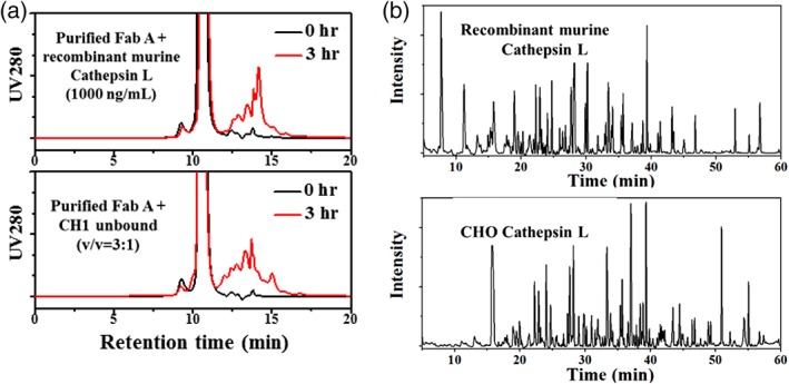
Figure 3.

Confirmation of Cathepsin L as the root cause of the fragmentation. (A) Fragmentation visualized by HP‐SEC profiles (during pH 3.4 incubation) for Fab A cleaved by recombinant murine Cathepsin L or CHO Cathepsin L (in CH1 unbound fraction). Fab A concentration was 2.5 mg/mL and 1 mM DTT was added in the sample mixture for the recombinant murine Cathepsin L. (B) Intact mass spectroscopy ion chromatograms for Fab A fragments generated by recombinant mouse Cathepsin L or CHO Cathepsin L (in CH1 unbound fraction).
