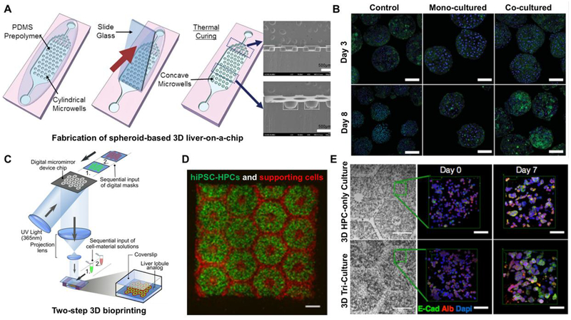
Figure 2.
Schematic showing microwell array PDMS plate based liver-on-a-chip device. (B) Generated 3D spheroids (mono-culture and co-cultured) on day 3 and 8. Reproduced from Lee et al (58) with permission from The Royal Society of Chemistry. (C) Hydrogel-based 3D bioprinted hepatic construct. hiPSC-HPCs and the support cells were patterned using two-step 3D bioprinting technique. (D) Fluorescent image is showing patterns of hiPSC-HPCs (green) and supporting cells (red). (E) Fluorescent images of albumin, E-cadherin, and nucleus staining of hiPSC-HPCs without supporting cells and in 3D triculture constructs. Reproduced from Ma et al. (63) with permission from Proceedings of the National Academy of Sciences of the United States of America.

