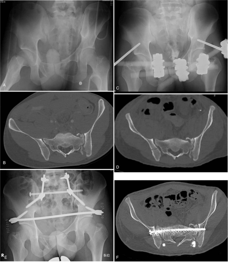
Figure 3.

Male, 26-years-old, crush injury. A. Radiograph on admission showing a vertically displaced sacral fracture on left. B. Axial CT view of S1 on admission showing a crescent fracture on right. C. Radiograph taken after external fixation. D. Axial CT view of S1 after external fixation. The sacral fracture on the left side is reduced. E. Post-operative radiograph: posterior pelvic ring was stabilized by MITO, and anterior subcutaneous internal fixation was applied. F. Post-operative axial CT view of S1 showing appropriate insertion of a trans-sacral screw. G. Iliac screws are not bulging from the surface of posterior superior iliac crests. CT = computed tomography, MITO = minimally invasive triangular osteosynthesis. H. Radiograph at 12 months post-injury. Implants were removed.
