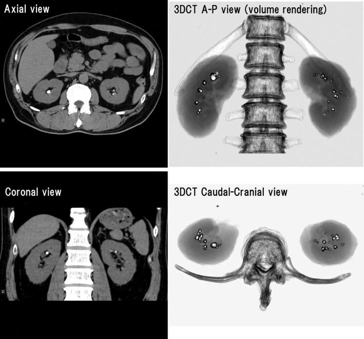
Figure 2.

Computed tomography (CT) scans of a 55‐year‐old man with gout since the age of 30 years. Multiple bilateral stones can be seen on the axial and coronal scans. Three‐dimensional CT (3DCT) was useful for confirming the distribution and number of stones, because stones in both kidneys could be easily observed on one screen with a high degree of accuracy
