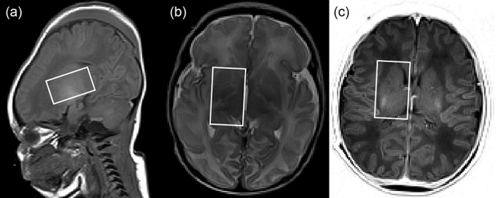
Figure 1.

Visualization of VOI. VOI with size of 14 ml (20 mm RL, 35 mm AP, 20 mm SI) is shown in A: sagittal T1‐weighted slice at the center position; B: transverse T2‐weighted slice at most inferior position; C: transverse IR‐weighted slice at the most superior position. In this infant the contribution of CSF to the VOI was 4.9%.
