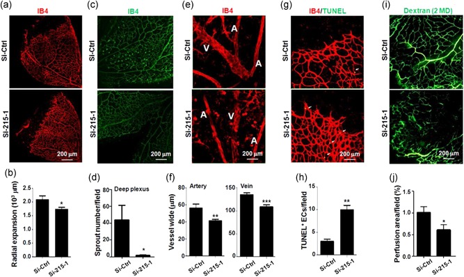
Figure 5.

Knockdown of TMEM215 resulted in abnormal retinal vessel development. P3 pups were intravitreally injected with chemically modified Si‐Ctrl or Si‐215‐1. Retinas were dissected on P7 and subjected to whole‐mount immunofluorescence staining. (a, b) Retinas were stained by IB4 (a), and diameter of retinal plexus was measured and compared (b) (n = 7). (c, d) Retinas were stained by IB4 (g), and deep plexus sprouting was counted (h) (n = 3). (e, f) Retinas were stained by IB4 (e), and width of arteries (a) and veins (V) was measured and compared (f) (n = 3). (g, h) Retinas were stained with IB4 and TUNEL (c), and apoptotic ECs were compared (d) (n = 3). (i, j) P3 pups were intravitreally injected with chemically modified Si‐Ctrl or Si‐215‐1. On P7, pups were injected with Dextran (MW, 2 MD) through left ventricle. Retinas were removed 10 min after the injection and photographed. Vessel perfusion was determined (j) (n = 3). Bar = means ± SD; *p < 0.05, **p < 0.01, and ***p < 0.001. EC: endothelial cells; IB4: isolectin B4; MW: molecular weight; TMEM: transmembrane protein; TUNEL: terminal deoxynucleotidyltransferase‐mediated dUTP nick end labeling; SD: standard deviation [Color figure can be viewed at wileyonlinelibrary.com]
