Figure 2.

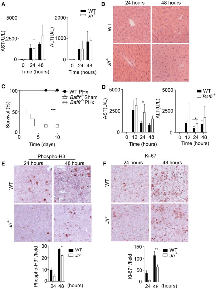
B cells play a crucial role in liver regeneration after PHx. (A) The activity of AST and ALT was measured in serum of WT and Jh–/– mice following PHx at the indicated time points (n = 4‐5). (B) Sections of snap‐frozen liver tissue from WT and Jh–/– mice following PHx were stained with H&E. One representative set of n = 3 is shown (scale bar, 200 μm). (C) Survival of Baffr–/– mice (n = 18) after 70% PHx compared to sham‐operated Baffr–/– mice (n = 4) and WT mice after 70% PHx (n = 7) was monitored. (D) The activity of AST and ALT was measured in serum of WT and Baffr–/– mice following PHx at the indicated time points (n = 4‐5). (E,F) Sections of snap‐frozen liver tissue from WT and Jh–/– mice following PHx were stained with (E) anti‐phospho‐H3 and (F) anti‐Ki‐67 antibodies. Representative sections for each time point are shown (n = 3; scale bar, 100 μm). Lower panels indicate quantification. Error bars in all experiments represent SEM; *P < 0.05, **P < 0.01, ***P < 0.001.
