Figure 4.

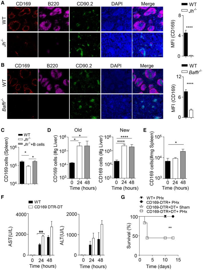
CD169+ cells contribute to liver regeneration. (A) Sections of snap‐frozen spleen tissue from WT and Jh–/– mice were stained with anti‐CD169, anti‐B220, and anti‐CD90.2 antibodies. Representative sections are shown (n = 4‐5; scale bar, 100 μm). Right panel indicates average and SEM of mean fluorescence intensities of CD169 staining. (B) Sections from snap‐frozen spleen tissue from WT and Baffr–/– mice were stained with anti‐CD169 anti‐B220 and anti‐CD90.2 antibodies. Representative sections are shown (n = 4‐5; scale bar, 100 μm). Right panel indicates average and SEM of mean fluorescence intensities of CD169 staining. (C) Purified B cells (2 × 10⁶) from WT mice were adoptively transferred into Jh–/– mice. After 48 hours, CD169‐cell numbers were determined in the spleen by flow cytometry (n = 4‐6). (D,E) CD169+ cells were measured by flow cytometry in the newly regenerated (n = 7‐8) and remaining (“Old”) (n = 3‐4) liver lobes (D) and spleen tissue (n = 7‐8) (E) at the indicated time points after 70% PHx. Results were calculated according to liver (grams) and spleen (milligrams) weights. (F) The activity of AST and ALT was measured in serum of WT and DT‐treated CD169‐DTR mice following PHx at the indicated time points (n = 3). (G) Survival of DT‐treated CD169‐DTR mice after PHx (n = 12) compared to WT mice (n = 8) after PHx, CD169‐DTR mice (n = 5) after PHx, and sham‐operated, DT‐treated CD169‐DTR mice (n = 4) was monitored. Error bars in all experiments represent SEM; *P < 0.05, **P < 0.01, ****P < 0.0001. Abbreviations: DAPI, 4′,6‐diamidino‐2‐phenylindole; MFI, mean fluorescence intensity.
