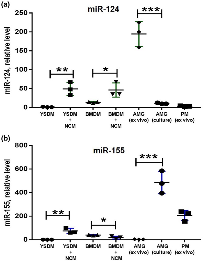
Figure 2.

Comparison of expression of miR‐124 and miR‐155 in bone marrow‐ and yolk sac‐derived macrophages under the influence of neuronal conditioned media. Bone marrow‐derived macrophages (BMDMs), yolk sac‐derived macrophages (YSDMs), or adult microglia were grown in the presence of M‐CSF as described in Materials and Methods. BMDMs and YSDMs were incubated in 50% neurobasal media (YSDM and BMDM) or 50% NCM (YSDM + NCM and BMDM + NCM) for 4 hr, and the expression levels of miR‐124 (a) and miR‐155 (b) were analyzed by real‐time RT PCR as described in Materials and Methods. The level of expression of miR‐124 and miR‐155 in YSDMs and BMDMs was compared to ex‐vivo isolated (AGM (ex vivo)) and cultured (AGM(culture)) adult microglia and ex‐vivo isolated peritoneal macrophages (PM (ex vivo)). In (a, b), mean ± SD of three separate experiments is shown on dotplots (n = 3; *p < 0.5; **p < 0.01; ***p < 0.001 as determined by unpaired Student's t test) [Colour figure can be viewed at wileyonlinelibrary.com]
