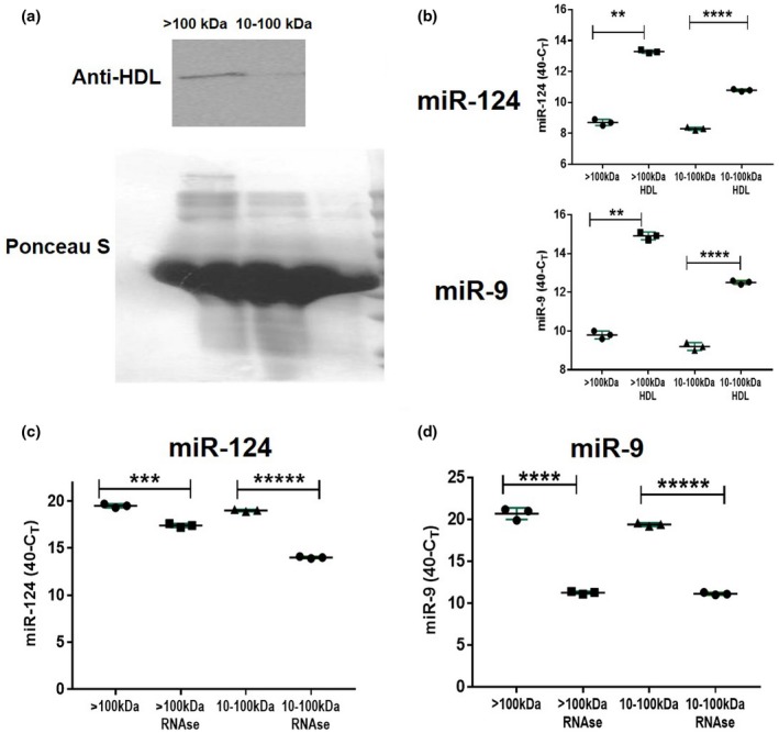
Figure 5.

Analysis of the level of expression of neuronal microRNAs miR‐124 and miR‐9 in HDL complexes. (a) Western blot analysis of the expression of 40‐kDa protein from HDL complexes in high molecular weight (>100 kDa) versus low molecular weight (10–100 kDa) fractions of BSCM. Ponseus S staining was used as a protein loading control. (b) Immunoprecipitation of HDL with miR‐124 and miR‐9. Anti‐HDL antibodies were adsorbed to plastic and >100‐kDa or 10–100‐kDa fractions of BSCM were added to antibodies as described in Materials and Methods. Expression of miR‐124 was assessed in HDLs bound to anti‐HDL antibodies (>100 kDa:HDL and 10–100 kDa:HDL) and in solution unbound to HDL in the >100 and 10–100‐kDa fractions (>100 and 10–100 kDa). (c, d) Comparison of expression of miR‐124 (c) miR‐9 (d) in untreated BSCM versus BSCM treated with RNase (fractions of >100 and 10–100 kDa). In (b–d), mean ± SD of three separate experiments is shown on dotplots (n = 3; **p < 0.01; ***p < 0.001; ****p < 0.0001; ****p < 0.00001; unpaired Student's t test) [Colour figure can be viewed at wileyonlinelibrary.com]
