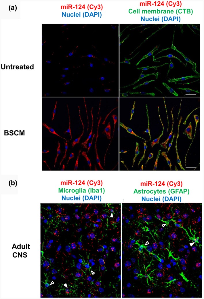
Figure 9.

Visualization of miR‐124 in macrophages in vitro and in microglia and astrocytes in vivo in mouse adult brain. Bone marrow‐derived macrophages (BMDMs) were incubated with medium (untreated) or in 50% brain slice conditioned medium (BSCM) for 4 hr as described in Materials and Methods. The cells were stained for cell plasma membrane marker GM1 with CTB‐FITC (green) and nuclei with DAPI (blue) as described in Materials and Methods. MiR‐124 (red) was detected by fluorescence in situ hybridization as described in Materials and Methods. (a) Histology sections were prepared from the normal adult brain of perfused 8‐ to 12‐week‐old B6 female mice as described in Materials and Methods. Microglia were stained with Iba1 (green; left image), and astroglia were stained with the anti‐GFAP antibody (green; right image). MiR‐124 (red) was detected by in situ hybridization as described in Materials and Methods. Open arrows indicated microglia and astrocytes without miR‐124 hybridization signal. Filled arrows indicate miR124‐positive microglia (left image) or astroglia (right image). Nuclei were stained with DAPI (blue). The bar is 10 μm
