
Figure 4.
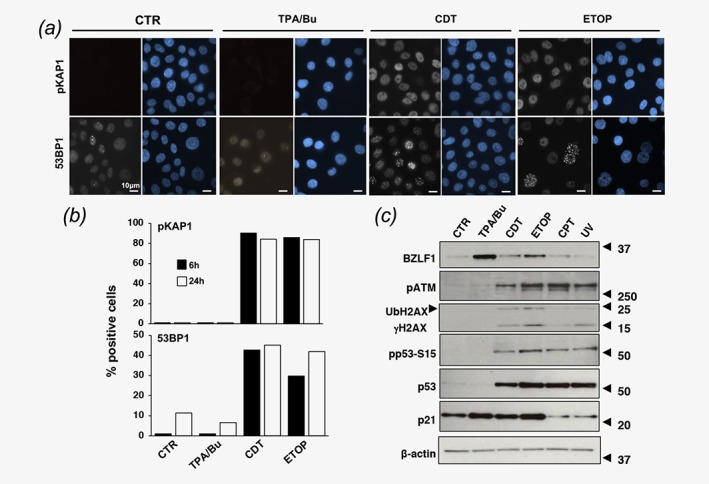
DNA double strand breaks promote EBV reactivation. (a). AGS‐Bx1 cells were exposed to TPA/Bu, CDT (1 μg/ml) or Etoposide (ETOP, 40 μM) for 6 hr or 24 hr. Activation of the DDR was monitored by detection of phosphorylated KAP1 (pKAP1, early marker) and 53BP1 foci (late marker). Nuclei were counterstained with DAPI (blue). Magnification 63 ×. (b). The percentage of positive cells assessed by scoring 90 cells per condition and time point. Cells with ≥5 53BP1 foci/nucleus were scored as positive. (c) AGS‐Bx1 cells were left untreated (CTR) or treated for 6 hr with TPA/Bu, CDT (1 μg/ml), Etoposide (ETOP, 40 μM), Camptothecin (CPT, 5 μM) or exposed to UV, and further incubated for 6 hr. Reactivation of the lytic cycle and induction of the DDR response were monitored by western blot using antibodies to BZLF1, phosphorylated ATM (pATM), γH2AX, p53, Ser15 phosphorylated p53 (pp53‐S15), and p21. The arrow indicates the ubiquitinated form of γH2AX (UbH2AX). β‐actin was used as loading control. One representative experiment out of three where all the antibodies were tested in parallel is shown.