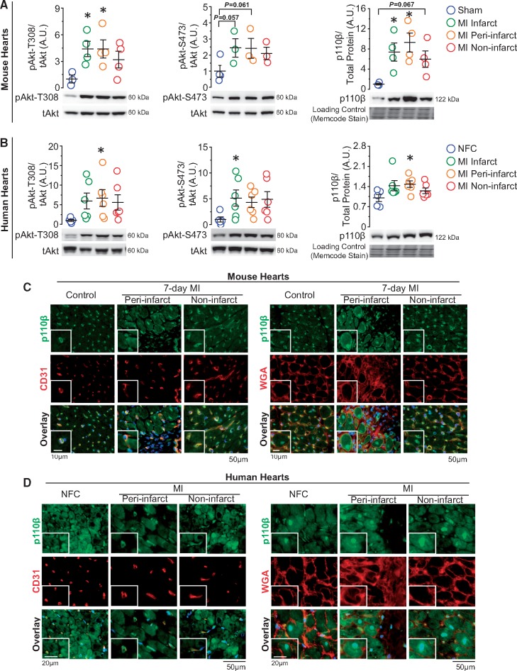
Figure 1.
Catalytic isoform of PI3Kβ-p110β is increased in post-MI murine and human hearts and expressed both in ECs and CMs. (A and B) Western blot analysis of Akt and p110β levels on 7-day post-sham/MI mouse hearts and on non-failing control (NFC) and post-MI failing human hearts. *P < 0.05 vs. sham/NFC hearts, n = 3–6 hearts/group (one-way ANOVA). (C and D) Immunofluorescence images of p110β (green) in the heart with EC marker-CD31 (red, left panels), WGA outlining CMs (red, right panels), and DAPI marking nuclei (blue) on mouse and human hearts, n = 3–4 hearts/group.

