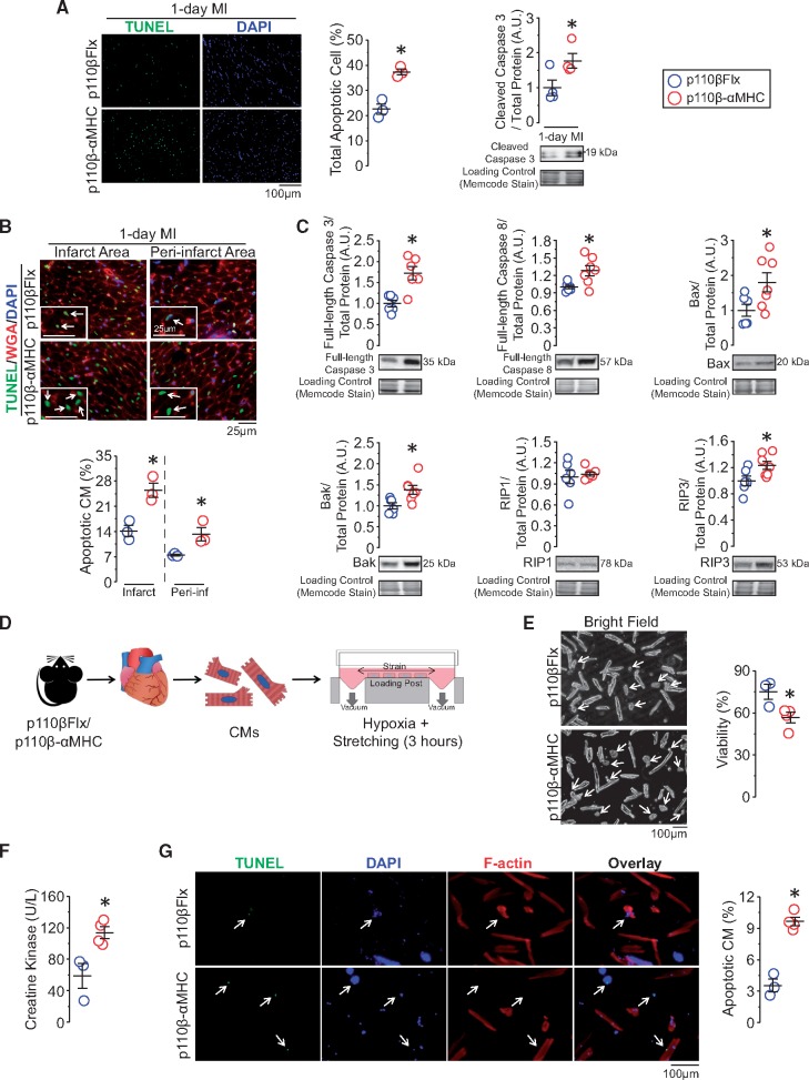
Figure 5.
Inactivation of CM-p110β sensitizes CMs to cell death by increasing pro-cell death proteins expression, leading to increased post-MI cell death. (A) Terminal deoxynucleotidyl transferase-mediated dUTP nick-end labelling (TUNEL, green) and DAPI (blue) immunofluorescence analysis for apoptotic cells (n = 3 mice/group) and western blot analysis for cleaved caspase 3 (n = 4 mice/group) on 1-day post-MI hearts. *P < 0.05 (t-test). (B) Combined wheat Germ Agglutinin (WGA, red), TUNEL, and DAPI immunofluorescence staining to highlight apoptotic CMs. *P < 0.05, n = 3 mice/group (t-test). (C) Western blot analysis for baseline protein levels of full-length caspase 3, full-length caspase 8, Bax, Bak, RIP1, and RIP3 in left ventricular lysates from p110β-αMHC and control mice. *P < 0.05, n = 6–7 mice/group (t-test). (D) Study design for isolated adult CM stretching under hypoxic condition for 3 h, n = 3–4 experiments/group (two hearts/experiment). (E) Representative bright field images and cell viability evaluation after stretching. *P < 0.05 (t-test). (F) Evaluation of creatine kinase level in the media from cultured CMs. *P < 0.05 (t-test). (G) TUNEL staining for apoptotic CMs (green) with F-actin (red) staining. *P < 0.05 (t-test).

