
Figure 3.
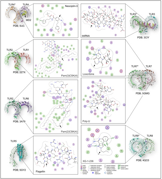
TLRs with bound ligands. The ligand binding mechanism of the extracellular TLRs (left, TLR1, 2, 4, 5, 6) and endosomal TLRs (right, TLR3, 7, 8) has been presented. Each monomer has been labeled; however, for the homodimers, the other monomer has been labeled with asterisk (*). In the case of TLR5, flagellin‐bound single monomer has been given. The respective protein databank (PDB) ID has also been given at the bottom of each structure. TLRs recognize several molecules, including protein, lipopeptide, small molecules and nucleic acids, and the bound ligand with individual TLR has been shown in 2D interaction diagram. The color code for the 2D interaction is given at the bottom of the figure. Black arrows indicate the bound TLR‐ligand. dsRNA, double‐stranded RNA; TLR, Toll‐like receptor; 2D, two‐dimensional [Color figure can be viewed at wileyonlinelibrary.com]