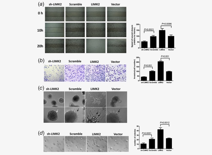
Figure 2.

Effect of LIMK2 expression level on UM‐UC‐3 bladder cancer cell metastasis potential. Cells were stably transfected with LIMK2‐targeted shRNA (sh‐LIMK2), scrambled control siRNA (Scramble), LIMK2 overexpression vector (LIMK2) or empty vector (Vector). (a) Wound healing assay. UM‐UC‐3 cells invasion was analyzed by video microscopy. Left panel: representative pictures of wound closure. Right panel: Speed of wound closure relative to empty vector (mean ± S.D). (b) Invasion assay. The invasive properties were analyzed in a Boyden chamber coated with Matrigel. (Left) Cells that adhered to the lower surface of the filter are shown. (Right) Results presented as mean cell number ± S.D. (c) Representative micrographs of cultured cells after 8 days of culture in 3D spheroid invasion assays. (d). Anchorage‐independent growth assay. Colonies >0.1 mm in diameter were counted under a microscopic field (Left). Results presented as mean ± S.D. (Right).
