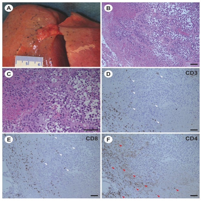
FIGURE 3.
Pathology evidence of antitumour response. (A) Small subpleural nodule of residual tumour in the left upper lobe of the lung. Similar nodules were found in the right upper and lower lobes. (B,C) Representative images of histology sections of the tumour nodule in the left upper lobe showing morphologic features of an epithelioid cell type melanoma (hematoxylin and eosin stain). The morphology of the tumour was similar in a premortem biopsy, when the tumour immunophenotype was determined. (D–F) Immunoperoxidase staining of serial sections of the left upper lobe tumour. T Cells positive for CD8 are located at the periphery and within the tumour nodule (white arrowheads); CD4-positive T cells are found at the periphery (red arrowheads). Magnification bars in all panels represent 100 μm.

