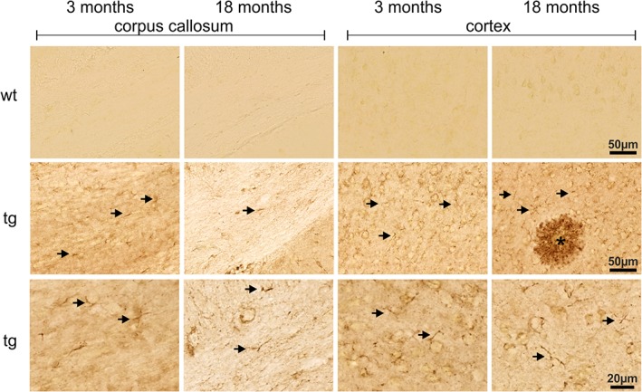
Figure 1.

Immunohistochemical detection of hAPP in corpus callosum and cortex of young and aged Tg2576 mice (tg) and wild type littermates (wt) as indicated. Immunoreactivity for hAPP is absent in wild type brain sections demonstrating the specificity of the 1D1 antibody. Although the majority of the 1D1 labeling arises from neurons, glial structures (arrows) are also hAPP‐immunoreactive. This is displayed at higher magnification in the bottom images (arrows)
