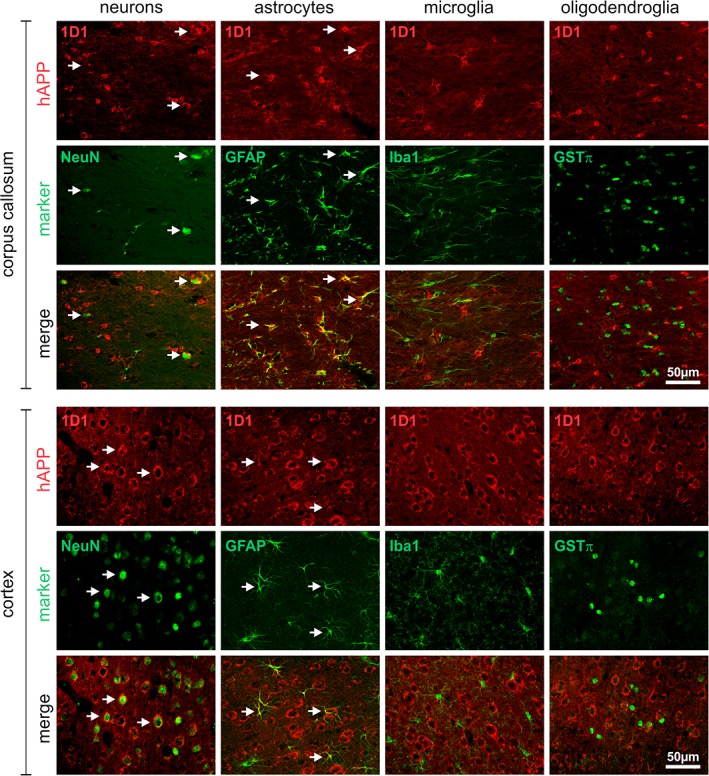
Figure 2.

Cell type‐specific expression of hAPP in brains of 3‐month‐old Tg2576 mice. The hAPP in corpus callosum and cortex was visualized using the antibody 1D1 and detection with secondary Cy3‐conjugated antibodies (red fluorescence) in combination with marker proteins for neurons (NeuN), astrocytes (GFAP), microglia (Iba1), and oligodendrocytes (GSTπ) detected with Cy2‐conjugated secondary antibodies (green fluorescence). Note the frequent co‐localization of hAPP with neurons and astrocytes (arrows)
