Figure 4.

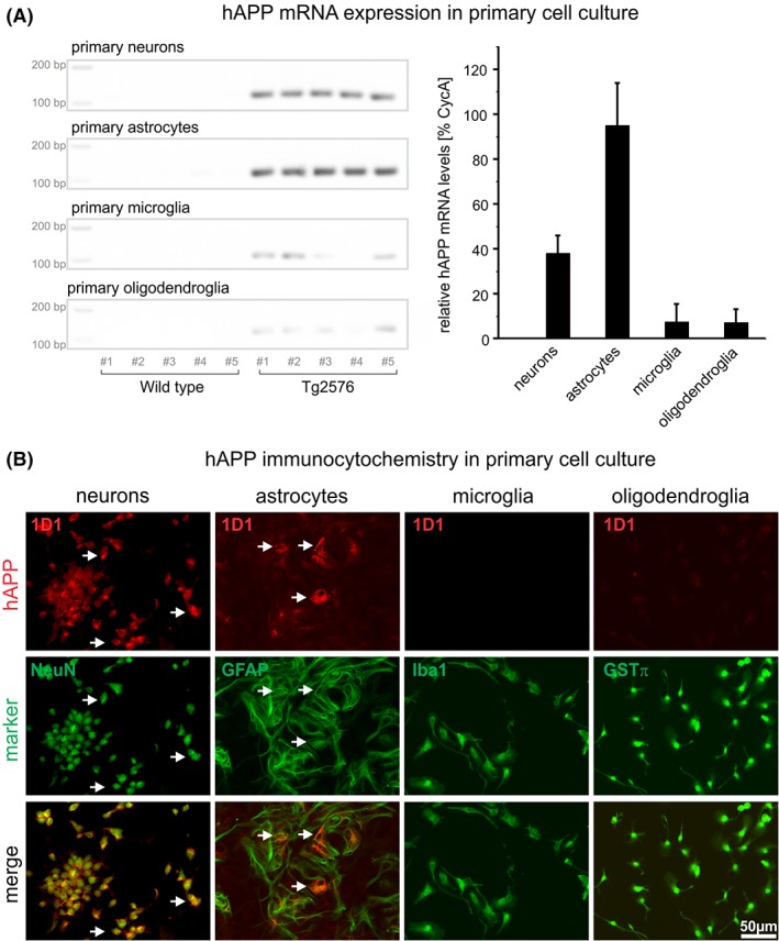
Specific detection of hAPP mRNA (A) and protein (B) in Tg2576 primary neurons and glial cells by RT‐qPCR and immunocytochemistry, respectively. (A) In neuronal and astrocytic and to a much lesser extent in microglial and oligodendroglial cultures of Tg2576 mice hAPP mRNA is detected, whereas in the corresponding cultures of wild type mice no hAPP mRNA was present. This is consistent with the specificity of primer pairs used for hAPP versus mouse APP. On the left hAPP PCR products separated on agarose gels from different cell types are shown. The diagram on the right shows the CycA‐normalized quantification of hAPP mRNA by RT‐qPCR in the respective cell types. (B) Primary neuronal and astrocytic cultures of Tg2576 mice display hAPP immunoreactivity (arrows), which is absent from microglial and oligodendroglial Tg2576 cultures
