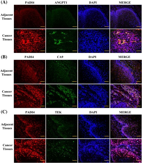
Figure 7.

Immunohistochemistry was used to detect the expression of PADI4 (red signals stained with goat anti‐mouse Alexa 568 secondary antibody) and ANGPT1, CA9, and TEK (green signals stained with goat anti‐rabbit Alexa 488 secondary antibody) in esophagus squamous cell carcinoma tissues and adjacent tissues. DAPI was used to stain the cell nuclei. A, The ANGPT1 expression level in cancer tissues and adjacent tissues. B, The CA9 expression level in cancer tissues and adjacent tissues. C, The TEK expression level in cancer tissues and adjacent tissues. Bar, 50 µm
