
Figure 2.
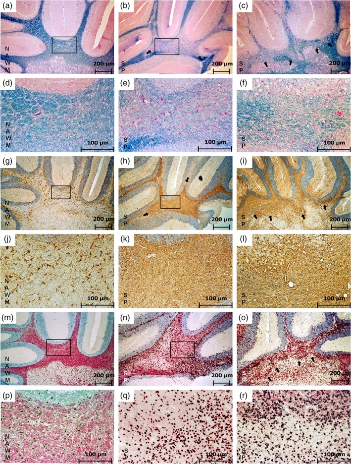
Shadow plaques in the rat brain. This figure shows a direct comparison of SP features and NAWM as found in the cerebellar white matter. Panels (a–f) represent LFB staining, (g–l) show GFAP‐IHC, (m–r) show PLP ISH. In NAWM (a, d) myelin appears in dark blue in LFB staining (a). The rectangle indicates the area represented in (d) at a higher magnification. The fully remyelinated SP appears in light blue due to thinner myelin sheaths after accomplished remyelination (b, c, e, f). The rectangle in (b) indicates the area shown in (e) and the arrows in (b) and (c) point at the SP border. Only some astrocytes appear GFAP positive in NAWM (g). The rectangle indicates the area represented in (j) at a higher magnification. In SP areas, astrocytes exhibit all features associated with a glial scar (h, i, k, l). The rectangle in (h) indicates the area represented in (k). The arrows in (h) and (i) point at the border of the glial scar. In NAWM, a normal occurrence of OPCs (black) and PLP (pink) can be observed (m). The rectangle indicates the area represented in (p) with a higher magnification. The distribution of OPCs in SP appears similar to their distribution in NAWM but with less PLP immunoreactivity in the background represented by a much paler pink background (n, o, q, r). The rectangle in (n) indicates the area shown in (q) and the arrows in (o) point to the SP border (magnification: (a–c), (g–i), (m–o) 50×; (d–f), (j–l), (p–r) 200×) [Color figure can be viewed at wileyonlinelibrary.com]