
Figure 4.
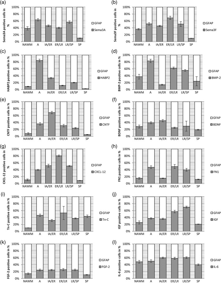
Percentage scale of GFAP positive astrocytes and astrocyte‐derived factors on selected spinal cord lesions. In each lesion type the mean of all astrocytes represents the 100% scale and the corresponding astrocytes expressing factors involved in OPC regulation are given in the appropriate ratios (for each factor n = 20 selected lesions per type). Error bars represent 95% CI. The guidance molecule Sema3A (a) peaked in A with more than 60% of active astrocytes expressing this factor and additionally showed a high occurrence in LR/SP with approximately 55%. In A, the expression of Sema3F (b) is higher than in IA/ER and peaks again in ER/LR. HABP2 (c) is highly present during A with more than 80% of all astrocytes expressing this factor. It decreases again to 5–30% during remyelination. BMP‐2 (d) is comparably high during A decreases in IA/ER and increases again during later remyelination steps. CNTF expression (e) increases during lesion evolution with a peak of almost 70% CNTF positive astrocytes in IA/ER and decreases again to approximately 5% in SP. BDNF (f) expression increases from NAWM to IA/ER, decreases in ER/LR, increases again in LR/SP and reaches its minimum in SP. CXCL‐12 (g) increases during lesion evolution, peaks in ER/LR with 80% of active astrocytes expressing this factor and decreases again from LR/SP to SP. Around 50% of all astrocytes are FN1 positive during A and late remyelination steps (h). Tn‐C is upregulated during A and ER/LR (i). The expression of IGF (j) rises from NAWM to LR/SP with a slight increase in A and falls rapidly from LR/SP to SP. FGF‐2 (k) showed only a slight increase from NAWM to A and decreases from LR/SP to SP. IL‐6 (l) shows a more uniform course with a slight increase of IL‐6 expression during remyelination process with more than 50% of all active astrocytes expressing IL‐6