
Figure 1.
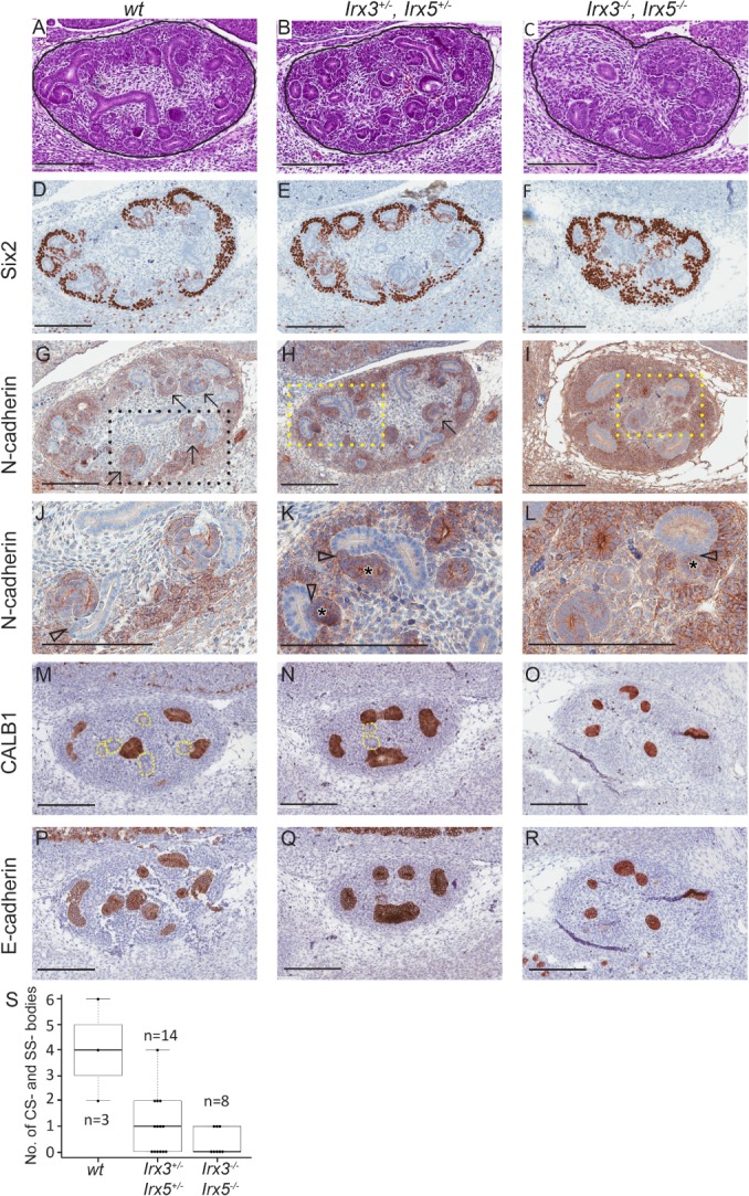
Reduced nephron morphogenesis in Irx3/Irx5 knockout mice. Representative images of Irx3 and Irx5 wt, heterozygous and homozygous knockout E13.5 foetal mouse kidney (FMK) sections stained with H&E (Htx; A–C). The CM/committed blastema, identified by Six2 expression was retained in all genotypes (D–F), although it was more extensively manifested in homozygous (Irx3 −/− Irx5 −/−) mice (F). Irx3/5 −/− mice, as opposed to wt and heterozygous mice, showed N‐cadherin positivity largely confined to cells of the CM as if differentiation through CSB and SSB had ceased (G–L). Specific labelling of UB structures with CALB1 (M–O), in combination with staining for all epithelial elements in the foetal kidney by E‐cadherin (P–R), confirmed that almost all epithelial structures were UB‐derived in Irx3/5 −/− mice (O and R). Irx3/Irx5 knockout mice maintained the ability to form some primitive nephrogenic tubules with a distinct lumen and could dock to UB‐derived structures, but the tails of the SSB were consistently missing in Irx3/Irx5 hetero‐ and homozygous knockout embryos, resulting in short tubules without curvature (K and L). Solid black lines demarcate the developing kidney from surrounding tissue (A–C). Arrows point to CSB and SSB in G and H. Content in rectangles with dashed lines in G–I are enlarged in J–L. Arrowheads point at docking sites between nephrogenic and UB‐derived epithelial structures, and asterisks (*) denote the lumen of nephrogenic tubules in K–L. Dotted lines in M and N correspond to E‐cadherin‐positive structures in P and Q. Scale bars correspond to 200 μm. All immunostains are brown. Box plot showing the median number and quartiles of CSB and SSB in FMK (S), where n denote the number of sections analysed for CSB and SCB in each group.