
Figure 4.
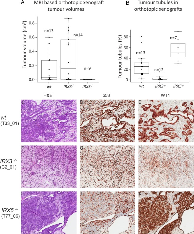
Loss of IRX3 promotes proliferative mesenchymal xenograft tumours, whereas loss of IRX5 stimulates differentiation of tumour tubules. (A) Tumour volumes based on MRI of NSG mice 12 weeks after xenograft transplantation with wt, IRX3 and IRX5 knockout orthotopic Wilms tumour xenografts. (B) The area of each tumour consisting of neoplastic tubules in wt, IRX3 and IRX5 knock out orthotopic xenografts. Boxplot represents median values and quartiles. (C–K): Detailed histological view of Wilms tumour orthotopic xenograft tumours with different IRX status, visualised by H&E staining (C, F, I) and IHC staining for p53 (D, G, J) and WT1 (E, H, K). The epithelial compartment of xenograft tumours was visualised by nuclear WT1 positivity, whereas p53 demarcated tumour cells from mouse host tissue. Within parentheses ( ) are the denotations of the specific xenograft tumours, listed in supplementary material, Table S3. Scale bar corresponds to 400 μm. All immunostains are brown.