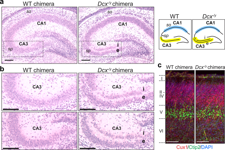
Figure 4. Dcx−/y NBC chimeras recapitulate the phenotype of germline Dcx−/y mice.
a, Representative images of H&E-stained coronal brain sections showing hippocampal defects in P0 Dcx−/y NBC chimeras (right) but not wild-type (WT) chimeras (left). Dashed lines highlight the CA3 region, where the stratum pyramidale (sp) is present as a single layer in WT chimeras (left) but is abnormally divided into an internal (i) and external (e) layer in the Dcx−/y chimeras (right). This phenotype is diagrammed on the right, with the CA3 (yellow) and CA1 (blue) regions highlighted. so, stratum oriens. b, Enlarged, representative images of the CA3 region in two P0 WT (left) and Dcx−/y (right) chimeras. c, Representative images of Cux1 (red), Ctip2 (green), and DAPI (blue) co-stained, coronal somatosensory cortex sections from P0 WT (left) and Dcx−/y (right) chimeras. Approximate location of cortical layers I-VI is indicated on the left. Scale bars, 100 μm. Images in a-c are representative of experiments performed on 21 mice (n=6 for Dcx−/y ESC clone 7F; n=4 for Dcx−/y ESC clone 8E; n=11 for wild-type ESCs). Mice were generated and characterized as depicted in Extended Data Figures 7–8.

