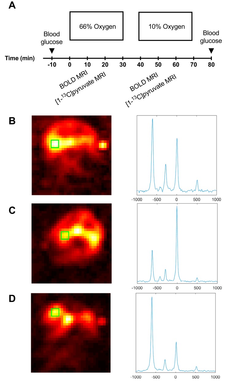
Figure 1.
Study protocol (A). All animals were exposed to either 66% oxygen content or 10% oxygen content in the breathing gas. Blood oxygen level–dependent (BOLD) scan is performed 10 minutes after the change of oxygen content followed by a hyperpolarized 13C pyruvate scan 20 minutes thereafter. Hyperpolarized 13C chemical shift imaging (CSI) image of a healthy control animal and accompanying kidney spectra from the green region of interest (ROI) (B). Hyperpolarized 13C CSI image of a untreated diabetic animal and accompanying kidney spectra from the green ROI (C). Hyperpolarized 13C CSI image of an insulin-treated diabetic animal and accompanying kidney spectra from the green ROI (D).

