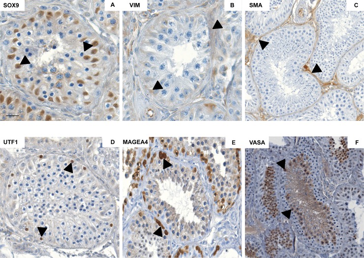
Fig 2. Characterization of somatic cell and germ cell markers in adult macaque testicular sections.
Representative images in the micrographs show strong expression for somatic cell markers SOX9 (A), VIM (B) and SMA (C) in adult macaque testicular sections. Intense expression of germ cell markers UTF1 (D), MAGEA4 (E) and VASA (F) was observed in adult macaque testicular sections in representative images in the lower panel. Black arrows in the micrographs indicate marker-specific localization. Scale bar in A, B, D and E indicates 50 μm, and in C and F indicates 100μm.

