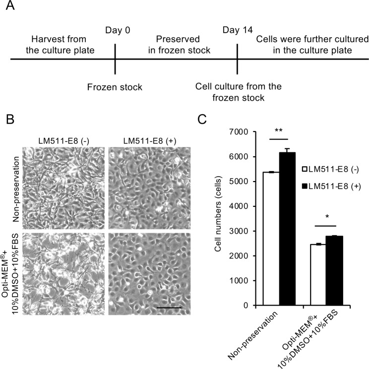
Fig 1. Cryopreservation of human corneal endothelial cells (HCECs) using Opti-MEM + 10%DMSO + 10%FBS.
(A) Time schedule for HCEC cryopreservation is shown. HCECs were harvested from the culture plate and suspended in cryopreservation reagent. After cryopreservation for 14 days, HCECs were seeded on the culture plate and cultured. (B) HCECs were suspended in Opti-MEM + 10%DMSO + 10%FBS, cryopreserved for 14 days, and seeded at the cell density of 6.0×10⁴ in one well of a 48-well culture plate for 24 hours. Three phase contrast images were obtained from each well of the 48-well culture plate, and randomly selected images by masked examiner are shown. Phase contrast microscopy showed fewer cells adhering to the culture plate, presumably due to the death of HCECs during preservation (left, lower), when compared to HCECs cultured without prior cryopreservation, shown as a control (left, upper). Inclusion of a laminin-511 E8 fragment increased the numbers of adhered HCECs. Scale bar: 200 μm. (C) Both the non-preserved and cryopreserved HCECs showed significantly increased cell numbers in response to the laminin-511 E8 fragment coating after 24 hours of culture. However, the cell numbers for the cryopreserved HCECs approximately half that obtained with the non-preserved HCECs. Experiments were performed in triplicate (n = 3). **p<0.01, *p<0.05.

