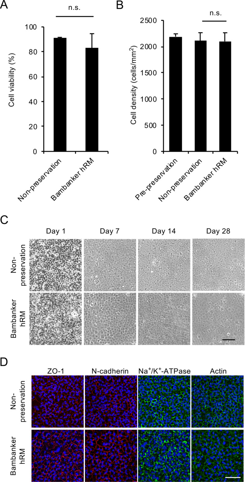
Fig 4. Feasibility of use of Bambanker hRM for preservation of clinical grade human corneal endothelial cells (HCECs).

(A) HCECs similar in grade to those used clinically (passage 3–5; cell density greater than 2000 cells/mm2) were cryopreserved using Bambanker hRM. The percentage of viable cells was 83.2% after 14 days of cryopreservation and 91.0% for a non-preserved control. Experiments were performed in duplicate. (B) HCECs were cultured for 28 days after cryopreservation, and cell density was evaluated by ImageJ. Cell density was not significantly decreased by cryopreservation when compared to non-preserved control cells (2099 cells/mm2 and 2111 cells/mm2, respectively). The statistical significance (vs. non-preservation) was determined with Student’s t-test (n = 4). (C) Phase contrast images showed that HCECs preserved with Bambanker hRM exhibited similar cell growth to that of a non-preserved control. Scale bar: 200μm. (D) HCECs cultured for 28 days after cryopreservation were evaluated for function-related markers by immunofluorescence staining. Actin distribution and cell morphology were evaluated by actin staining. HCECs preserved with Bambanker hRM showed similar expression of ZO-1, N-cadherin, and Na+/K+-ATPase at the cell-cell border to that seen in the non-preserved control. Actin was distributed at the cell cortex in the preserved HCECs and showed the normal corneal endothelial pattern seen in the non-preserved control. Scale bar: 200 μm.
