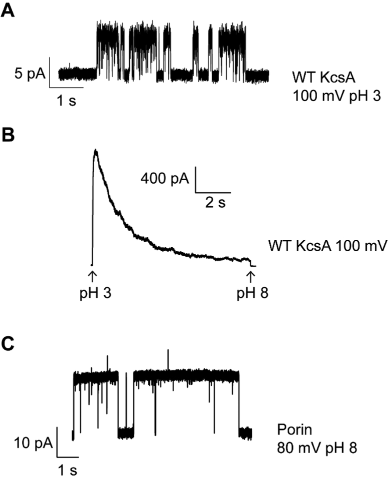
Figure 3.
Electrophysiological recordings of ion channels.(A) Single channel recording of KcsA at 100 mV with 200 mM KCl, 10 mM succinate pH 3.0 in the bath and 200 mM KCl, 0.5 mM CaCl2, 10 mM succinate-KOH pH 4.0 in the pipette. (B) Macroscopic recording of KcsA at 100 mV. Channels were activated by rapid perfusion of the bath solution from 200 mM KCl, 10 mM MOPS-KOH pH 8.0 to 200 mM KCl, 10 mM succinate pH 3.0. On the change in pH, the channels rapidly activate and then inactivate. (C) Representative porin single channel activity with 200 mM KCl in the recording solutions. Porins are a common contaminant that can easily incorporate into blisters.

