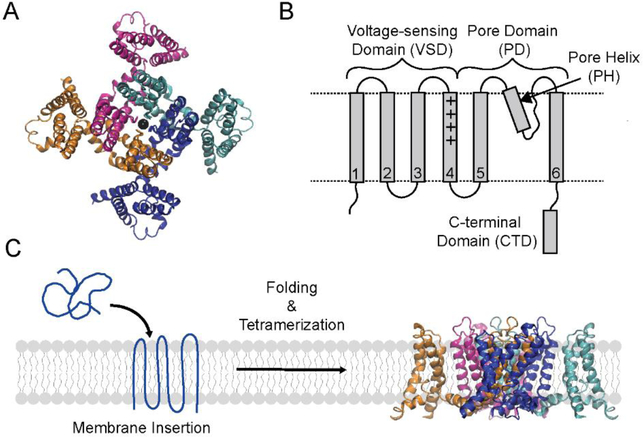
Figure 1. Structure and folding of the KvAP channel.
(A) Top view of the tetrameric KvAP channel. The structural model of the KvAP channel previously described31 is shown. The structure illustrates the domain swap between the pore and voltage sensor domains of adjacent subunits in the KvAP channel. (B) Topology map illustrating the domains and the structural features of a KvAP channel subunit. Dashed lines are membrane boundaries. (C) The folding pathway of the KvAP channel involves the stages of membrane insertion, secondary/tertiary folding and tetramerization. Structure figures were generated with VMD.60

