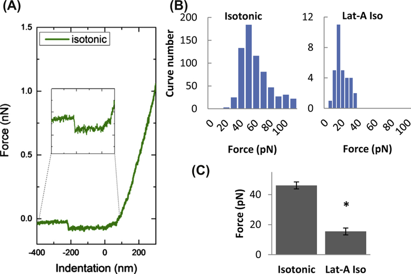
Figure 3.

Effect of F-actin depolymerization on the force of membrane tether formation. (A) Representative traces of AFM retraction force curves for cells in isotonic solution with an inset of a representative force discontinuity (used to obtain the tether force). The force discontinuity represents the change in force experienced by the cantilever while retracting from the sample surface upon detachment of the tether. (B) Histograms of membrane tether forces measured in cells exposed to isotonic solution (left) and Latrunculin-A in isotonic medium (right). (C) Mean membrane tether forces of cells with and without exposure to Latrunculin-A (1 µM for 10 min). (Asterisk denotes statistically significant difference using an unpaired samples T-Test (n = 15–60 cells; P < 0.05) between Latrunculin-A treated and untreated cells).
