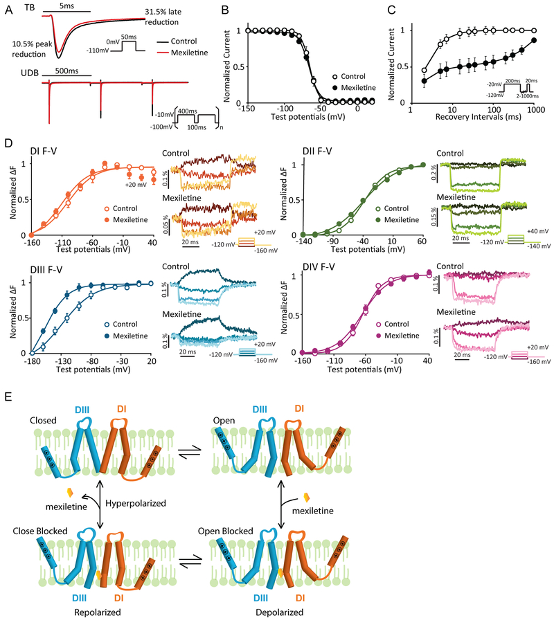
Figure 1: Mexiletine blockade of NaV1.5 channel stabilize the DIII-VSD at the activated position.
A. Representative current traces before and after 250 μM mexiletine tonic block (TB) and use-dependent block (UDB). Comparison between traces before and after mexiletine shows that mexiletine reduces the peak current by 10.5%, but the later component (10ms after peak) by 31.5%. 250 μM mexiletine was used, because channels exhibit moderate TB and UDB at this concentration.
B. Steady-state inactivation (SSI) curves before and after 2 mM mexiletine (n=4). Channel SSI was tested by holding the cells from −150 to 20 mV with a 10-mV increment for 200 ms. Fraction of channels available were then measured by peak currents induced by a −20mV test pulse. Mexiletine induces minimal hyperpolarizing shift in SSI curve.
C. Channel recovery from inactivation curves before and after 250 μM mexiletine (n=3). Cells were first depolarized to −20 mV to induce inactivation, then allowed to recover at −120 mV for various durations. Fraction of channels recovered were then tested with a −20mV pulse. Mexiletine slows down both phases of recovery, especially the slow recovery.
D. Left panels: Voltage dependence of steady-state fluorescence (F-V curves) from four domains (DI-V215C, DII-S805C, DIII-M1296C, DIV-S1618C) before and after 4 mM mexiletine. The mean ± SEM is reported for groups of 4 to 8 cells. Fluorescence after mexiletine was measured after 80% tonic block. Right panels: representative fluorescence traces before and after mexiletine. Four voltage steps ranging from −160 to 20mV (DI, DIII, and DIV) or −140 to 40mV (DII) at a 40mV interval are shown. Mexiletine only affects DIII-VSD by causing a hyperpolarizing shift in DIII F-V curve and slows down DIII-VSD deactivation, without affecting other three domains.
E. Proposed schematic (adapted from Arcisio-Miranda lidocaine model31) showing the mechanism of mexiletine stabilization of activated DIII-VSD. Only DI and DIII are shown and the VSDs are represented by a single S4 segment for clarity.

