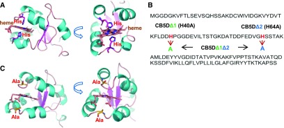
Figure 8.
Creation of CB5D Variants.
(A) Homology model of AtCB5D based on the crystal structure of human CB5B (PDB ID: 3NER). The bound heme and its interacting His residues are presented as sticks.
(B) Diagram of the substitutions (His to Ala) in AtCB5D. Mutants with substitutions of His-40, or His-64, or both with Ala are designated as CB5DΔ1, CB5DΔ2, and CB5DΔ1Δ2, respectively.
(C) Homology model of CB5DΔ1Δ2, showing its identical overall structure to the parental protein.

